Fusion Ultrasound Improving Outcomes in Liver Cancer Treatment
Treatment for liver cancer depends on the extent (stage) of the disease, your age, overall health and personal preferences. Operations used to treat liver cancer includes: open surgery to remove the tumor and minimally invasive local therapy. In minimally invasive local therapy, an interventional radiologist uses the advanced visualization technology in order to determine the lesion and perform local ablation. This reduces hospital stay and complications. This new imaging technique allows the doctor to conduct the procedure with ease and confidence.
Advances in the treatment of liver cancer
Ultrasonography is a type of imaging that uses high-frequency sound (ultrasound) waves to produce images of internal organs and other tissues. It’s effectively used to produce images of internal organs in the abdomen, pelvis, chest, and to guide doctors when they remove a sample of tissue for a biopsy or perform a procedure. Bone or gas can block ultrasonography, Thus using it to obtain images of certain structures is difficult. Also, there might be difficulties imaging structures deep in the body, especially in obese patients. In some patients, the doctor might not be able to perform a procedure such as microwave ablation of liver tumor, because the ultrasound image is not clear enough.
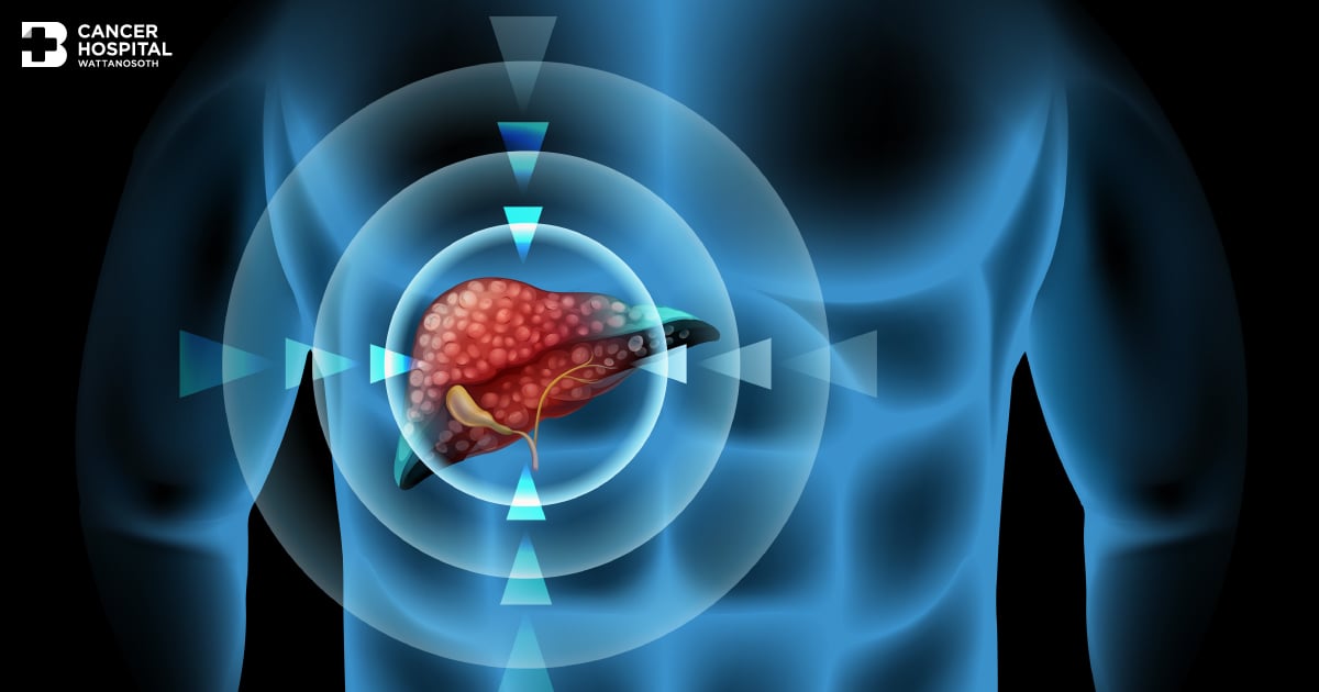
With technological advancements, there is a new imaging technique, called “fusion imaging”. This allows the doctor to fuse CT-scan/MRI images with live ultrasound so they can make confident decisions even in challenging diagnostic cases. Real-time fusion imaging can simultaneously fuse two different images and produce results in 4D image with increased accuracy. This allows the doctor to see internal organs and tumor clearly.
Assoc. Prof. Dr. Komgrit Tanisaro, Head of Interventional Radiology Center at Wattanosoth Hospital
, explained that this new fusion imaging allows the doctor to see the tumor in the liver clearly and precisely. In microwave ablation procedure, microwaves transmitted through the probe are used to heat and destroy the tumor. This can increase monitoring and targeting confidence during the procedure. Patients will receive an effective treatment that has low rate of complications and less recovery time. Most patients can go back to work in 2-3 days as the surgical wound is only 2-3 millimeters. Therefore, this treatment option increases survival rate and improves the patient’s quality of life.